
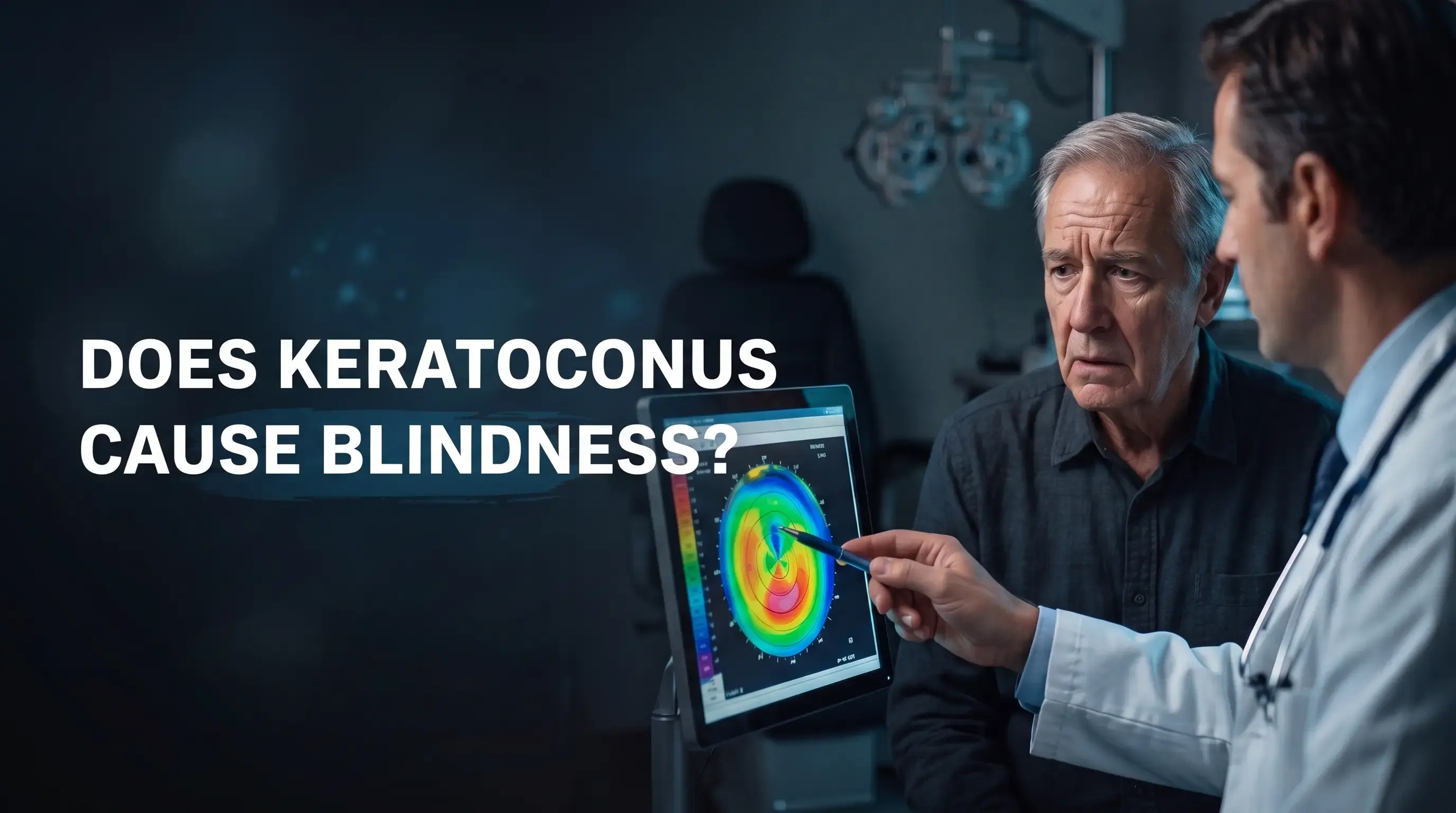
Quick answer: Keratoconus rarely causes total blindness. Keratoconus affects only the cornea, not the retina or optic nerve, so useful vision can almost always be restored. However, untreated progressive keratoconus can seriously reduce vision. Cross-linking can stop it from getting worse, rigid or scleral lenses can restore clear vision, and a corneal transplant can help in advanced cases.
Key Takeaways
- Keratoconus rarely causes total blindness, because it affects the cornea rather than the nerve of the eye.
- Without treatment, progressive keratoconus can cause severe blurred vision and scarring.
- Cross-linking aims to stop keratoconus from getting worse; it is often recommended at diagnosis in young patients.
- Rigid or scleral lenses give most patients clear vision; a corneal transplant helps in advanced cases.
- Early diagnosis is the best protection for your vision.
Can Keratoconus Make You Blind?
Keratoconus rarely causes total blindness. The cornea becomes thin and irregular, which blurs and distorts vision, but the light-sensing parts of the eye usually stay healthy. Even when vision with glasses is very poor, it can usually be improved with specialty contact lenses or a corneal transplant.
However, untreated progressive keratoconus can reduce vision enough to affect driving, reading and work. In advanced cases, the cornea can scar, and rarely it can suddenly swell (acute hydrops). That is why keratoconus should be monitored and treated early.
How Is Vision Protected and Restored in Keratoconus?
| Treatment | What it does |
|---|---|
| Corneal cross-linking | Aims to stop keratoconus from getting worse |
| Rigid and scleral lenses | Give clear vision by creating a smooth surface over the cornea |
| Corneal rings | Flatten the cornea to improve vision or lens comfort |
| Corneal transplant (usually DALK) | Replaces the damaged cornea in advanced cases with scarring |
Read about all keratoconus treatment options.
Why Does Early Diagnosis Matter?
Cross-linking can stop keratoconus from getting worse, but it cannot reverse damage that has already happened. Finding keratoconus early, often from rising astigmatism or a corneal scan before LASIK, gives the best chance of keeping good vision. Read about how keratoconus is diagnosed and the stages of keratoconus.
What Is Acute Hydrops?
Acute hydrops is a sudden swelling of the cornea in advanced keratoconus, when a small tear in the inner layer lets fluid into the cornea. It causes sudden blurred vision, pain, watering and a white patch on the cornea. It needs urgent care. It usually settles over weeks to months, sometimes leaving a scar, and does not usually cause blindness.
Can You Drive and Live Normally With Keratoconus?
Most people with keratoconus live normal lives, study, work and drive, especially with the right lenses. Driving depends on meeting the legal vision standard with your glasses or lenses. Glare at night can be a problem in more advanced keratoconus, so discuss it with your doctor.
What Can You Do to Protect Your Vision?
- Do not rub your eyes, and treat any eye allergy.
- Attend regular scans, especially in your teens and twenties.
- Have cross-linking if keratoconus is progressing.
- Ask your family to be screened.
- Do not have LASIK if you have keratoconus.
What Patients Say (Google Reviews)
My daughter’s experience with Dr. Fouad El Sayyad was very successful, thanks to God and then Dr. Fouad. It was a corneal cross-linking procedure, followed by the use of rigid contact lenses. … They are honest in how they deal with each case.
Google review · Sayyad Eye Center, Mohandeseen · October 2026 · translated from Arabic
My sincere thanks and appreciation to Dr. Fouad and Sayyad Center. I had a corneal transplant in my right eye and the operation was very successful, thanks to God and then Dr. Fouad. Four years later, after the success of the first operation, I had a transplant in my left eye, and thank God my vision has now improved a lot in both eyes.
Google review · Sayyad Eye Center, Mohandeseen · October 2026 · translated from Arabic
Where Does Dr. Fouad El Sayyad See Patients?
- Sayyad Eye Center, Mohandeseen: 3A Gameat El Dowal El Arabeya St., Sphinx Square.
- Sayyad Eye Center, New Cairo: Medical Park 2, opposite New Cairo Court.
To book an appointment with Dr. Fouad El Sayyad, call or WhatsApp 01000088168.
Frequently Asked Questions
Does keratoconus cause blindness?
Keratoconus rarely causes total blindness, because it affects the cornea rather than the retina or optic nerve. Untreated, it can seriously reduce vision, but useful vision can almost always be restored with lenses or a corneal transplant.
When is keratoconus dangerous?
Keratoconus threatens vision when it keeps progressing without treatment or reaches an advanced stage with scarring. Sudden pain with sudden blurring may be acute hydrops and needs urgent care.
Can keratoconus be cured?
Keratoconus cannot be reversed, but it can usually be controlled. Cross-linking aims to stop it from getting worse, and lenses, rings or a transplant can restore useful vision.
Can keratoconus be treated with laser?
LASIK is not done for keratoconus, because it can weaken the cornea further. Cross-linking uses ultraviolet light, not a cutting laser, to strengthen the cornea, and a femtosecond laser is used to create channels for corneal rings.
Can eye rubbing make keratoconus worse?
Yes. Frequent eye rubbing is strongly linked to keratoconus getting worse. Treat any itching with the drops your doctor prescribes and avoid rubbing.
Can a patient with keratoconus have LASIK?
No. LASIK can make keratoconus worse. In selected patients with stable keratoconus, an implantable contact lens (ICL) may be considered after a full assessment.
At what age does keratoconus stop progressing?
Keratoconus usually progresses most in the teens and twenties and often slows in the thirties, but this varies. That is why scans are repeated over time.
Can people with keratoconus drive?
Yes, as long as they meet the legal vision standard with their glasses or contact lenses. Rigid or scleral lenses often make this possible even when glasses are not enough.
This article is for general education and does not replace an eye examination. Treatment decisions depend on your own eye assessment.
References
This article is for general education and does not replace an eye examination. Treatment decisions depend on your own eye assessment.
FAQS
-
Does keratoconus cause blindness?
Keratoconus rarely causes total blindness, because it affects the cornea rather than the retina or optic nerve. Untreated, it can seriously reduce vision, but useful vision can almost always be restored with lenses or a corneal transplant.
-
When is keratoconus dangerous?
Keratoconus threatens vision when it keeps progressing without treatment or reaches an advanced stage with scarring. Sudden pain with sudden blurring may be acute hydrops and needs urgent care.
-
Can keratoconus be cured?
Keratoconus cannot be reversed, but it can usually be controlled. Cross-linking aims to stop it from getting worse, and lenses, rings or a transplant can restore useful vision.
-
Can keratoconus be treated with laser?
LASIK is not done for keratoconus, because it can weaken the cornea further. Cross-linking uses ultraviolet light, not a cutting laser, to strengthen the cornea, and a femtosecond laser is used to create channels for corneal rings.
-
Can eye rubbing make keratoconus worse?
Yes. Frequent eye rubbing is strongly linked to keratoconus getting worse. Treat any itching with the drops your doctor prescribes and avoid rubbing.
-
Can a patient with keratoconus have LASIK?
No. LASIK can make keratoconus worse. In selected patients with stable keratoconus, an implantable contact lens (ICL) may be considered after a full assessment.
-
At what age does keratoconus stop progressing?
Keratoconus usually progresses most in the teens and twenties and often slows in the thirties, but this varies. That is why scans are repeated over time.
-
Can people with keratoconus drive?
Yes, as long as they meet the legal vision standard with their glasses or contact lenses. Rigid or scleral lenses often make this possible even when glasses are not enough.
-
Can patients with keratoconus drive?
Many patients with keratoconus can drive if their corrected vision meets the required legal and safety standards. Some patients may need specialty contact lenses to achieve adequate vision.

Book an Eye Consultation Appointment | Dr. Fouad El Sayyad
Book now