
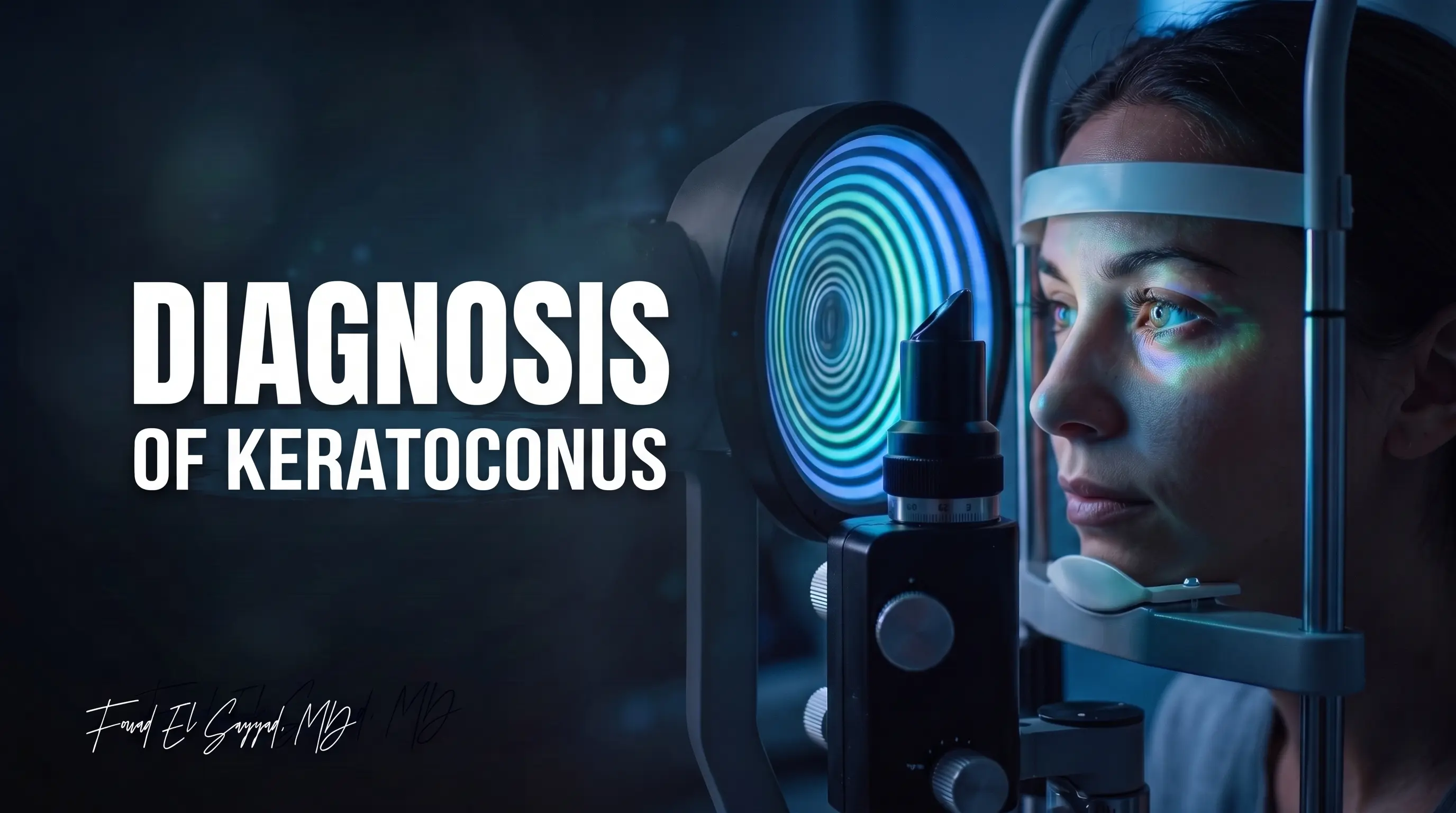
Diagnosis of keratoconus

Home Diagnosis of keratoconus

It may begin with mild blurred vision or the frequent need to change eyeglasses. However, in some cases, these signs may be an early indication of keratoconus. This is where the importance of accurate and timely keratoconus diagnosis becomes clear, as early detection plays a major role in preserving vision quality and reducing disease progression over time.

In this article, we will discuss the importance of keratoconus diagnosis and the most important examinations used to detect the disease, such as Corneal Topography, Pentacam examination, and Pachymetry. We will also explain the difference between keratoconus and regular astigmatism, and when the necessary tests should be performed.

In addition, we will explain how a patient with keratoconus sees, what the stages of keratoconus are, and whether the condition is considered serious. We will also highlight the latest diagnostic devices available at Dr. Fouad El-Sayyad Center and the importance of medical expertise in detecting early cases and creating a suitable treatment plan for keratoconus. Continue reading to the end to learn more.

Keratoconus Diagnosis

Keratoconus diagnosis is an essential step in the early treatment of this condition, which affects the cornea and causes it to take on an irregular shape.

Keratoconus is a condition in which the cornea changes from its normal round shape into a cone-like shape, affecting visual clarity. It is considered a progressive disease. When detected early, diagnosis can greatly improve the treatment approach and help limit vision deterioration.

At Dr. Fouad El-Sayyad Center, the latest medical tools are used to diagnose keratoconus accurately. With the advancement of keratoconus diagnostic techniques, it has become easier to detect the condition in its early stages using advanced examinations such as Corneal Topography and Pachymetry, which help determine the degree of corneal damage accurately.

Many patients can live a normal life after keratoconus treatment if diagnosis and treatment are performed at the right time. Therefore, it is important not to ignore any symptoms of keratoconus, such as persistent blurred vision or frequent changes in eyeglass prescription, and to visit a doctor when any of these symptoms appear.

When Should Keratoconus Diagnostic Tests Be Performed?

When experiencing any symptoms of keratoconus, such as blurred vision or increased astigmatism, the necessary tests should be performed to diagnose the condition.

In some cases, the symptoms may be very mild at first, making diagnosis difficult. However, over time, if early detection is delayed, the condition may worsen and affect vision more significantly.

When Should You Visit a Doctor for an Examination?

You should visit a doctor for keratoconus evaluation in the following cases:

  • Frequent changes in eyeglass prescription:
    If eyeglasses do not fully improve vision despite repeated prescription changes.

  • Blurred vision, especially at night:
    The patient may experience hazy vision or halos around lights at night.

  • Severe sensitivity to light:
    If you feel unusually sensitive to natural or artificial light.

  • Persistent headaches or eye strain:
    In some cases, continuous headaches may occur due to excessive visual effort caused by blurred vision.

If any of these symptoms appear, visiting the doctor should be a priority. Early keratoconus diagnosis can help choose the most suitable treatment decision, such as corneal cross-linking or the use of specialized contact lenses.

How Does a Patient with Keratoconus See?

The vision of a patient with keratoconus differs from normal vision because of the change in corneal shape and the irregularity of its surface. This causes light to scatter inside the eye instead of focusing clearly on the retina.

As a result, many patients experience gradual blurred vision that may worsen over time according to the stage of keratoconus and the degree of disease progression.

A patient may notice several changes in vision quality, such as:

  • Persistent blurry or hazy vision.

  • Seeing shadows or double images around objects.

  • Halos around lights, especially at night.

  • Difficulty driving at night due to light scattering.

  • Difference in vision quality between the two eyes.

  • Difficulty seeing fine details clearly, even with eyeglasses.

These symptoms gradually increase as the cornea changes shape and its irregular curvature becomes more severe. Therefore, the patient may feel that eyeglasses no longer provide the same quality of vision, even after changing the prescription more than once.

Specialized examinations such as corneal mapping and Pentacam help the doctor understand how a patient with keratoconus sees more accurately. They also help determine the degree of visual distortion caused by the disease, which supports choosing the most suitable treatment method to improve vision quality.

Steps of Clinical Eye Examination for Detecting Keratoconus

When visiting an experienced ophthalmologist such as Dr. Fouad El-Sayyad, Consultant of Ophthalmology and Eye Surgery and holder of the American Board certification, several steps are taken to diagnose keratoconus accurately.

The clinical examination depends on a group of tests that help the doctor evaluate eye health and determine the extent to which keratoconus affects vision.

Main Examinations Used at Dr. Fouad El-Sayyad Center to Diagnose Keratoconus

  • Slit-lamp examination:
    This allows the doctor to examine the eye thoroughly, including the front layers of the cornea, to detect any scars or abnormalities that may affect vision.

  • Corneal Topography:
    This test provides a three-dimensional image of the cornea and identifies irregular curvature on the corneal surface, helping detect the presence of keratoconus.

  • Pachymetry:
    This test measures corneal thickness and gives an idea of corneal strength, as thinner corneas may be more affected by keratoconus.

  • Keratometry:
    Corneal curvature is measured using a keratometer. This helps determine the degree of irregular corneal curvature.

After these examinations are performed, the doctor can determine the stage of keratoconus and create the most suitable treatment plan according to the results.

Role of Corneal Topography in Accurate Keratoconus Diagnosis

Corneal Topography is considered one of the most important examinations used in keratoconus diagnosis. It helps create a detailed map of the shape and curvature of the corneal surface with high accuracy.

This test is used to detect abnormal changes in corneal shape, even in early stages that may not appear during a traditional eye examination.

The examination works by analyzing light reflection on the corneal surface to produce a colored map that shows areas of normal and abnormal curvature. This helps the doctor evaluate the shape of the keratoconic cornea and determine the extent of disease progression.

Corneal Topography is also used to monitor disease progression over time and compare results between different visits.

The most important information provided by corneal imaging includes:

  • Accurate determination of corneal steepness.

  • Detection of irregular curvatures associated with keratoconus.

  • Evaluation of corneal symmetry between both eyes.

  • Monitoring disease progression over time.

  • Helping determine the most suitable keratoconus treatment options.

For this reason, Corneal Topography is considered one of the essential examinations that support early diagnosis and reduce the chances of vision deterioration over time.

Pentacam Examination and Its Importance in Detecting Early Cases

Pentacam examination is one of the most advanced tools used in keratoconus diagnosis. It provides a three-dimensional analysis of the cornea and the anterior segment of the eye with high accuracy.

This examination helps detect very early cases that may not appear clearly in traditional examinations.

The Pentacam device uses Rotating Scheimpflug Camera technology. It captures hundreds of points inside the cornea within a few seconds to produce accurate data about corneal shape, thickness, and anterior and posterior curvature.

The main advantages of Pentacam examination include:

  • Detecting keratoconus in very early stages.

  • Accurately analyzing the anterior and posterior surfaces of the cornea.

  • Measuring corneal thickness in all areas.

  • Evaluating disease progression and monitoring it over time.

  • Helping determine whether the cornea is suitable for vision correction surgeries.

The examination also helps the doctor better understand how a patient with keratoconus sees by analyzing visual distortions and optical aberrations caused by changes in corneal shape.

Therefore, the Pentacam device is an important tool in creating an accurate treatment plan for keratoconus according to each individual case.

Pachymetry and Its Effect on the Treatment Plan

Pachymetry, or corneal thickness measurement, is one of the essential examinations in keratoconus diagnosis. It helps accurately determine corneal thickness, especially because the cornea gradually becomes thinner as the disease progresses.

Corneal thickness directly affects the choice of the most suitable treatment plan for each patient.

Thickness is measured using specialized devices that work with ultrasound or optical imaging. These devices provide accurate measurements for different areas of the cornea, helping the doctor assess corneal stability and the severity of the condition.

The most important benefits of Pachymetry include:

  • Evaluating the degree of keratoconus progression.

  • Determining the safety of performing corneal cross-linking.

  • Assessing whether the cornea is suitable for vision correction surgeries.

  • Monitoring corneal thinning over time.

  • Helping choose the most appropriate keratoconus treatment methods.

This test also helps evaluate the stages of keratoconus and identify cases that may require early therapeutic intervention before greater vision deterioration occurs.

For this reason, Pachymetry is considered an important part of any accurate plan for keratoconus diagnosis and follow-up.

Difference Between Keratoconus Diagnosis and Regular Astigmatism

Some symptoms of keratoconus may be similar to regular astigmatism, such as blurred vision or the frequent need to change eyeglasses. Therefore, it may be difficult to distinguish between them in the early stages without accurate examinations.

The main difference is that regular astigmatism is often stable and regular, while keratoconus causes gradual and irregular changes in corneal shape, making vision more distorted over time.

In regular astigmatism cases, vision usually improves with glasses or traditional contact lenses. However, a patient with keratoconus may notice that glasses no longer provide clear vision even after changing the prescription several times.

The shape of the keratoconic cornea is also more irregular compared with normal astigmatism. This appears clearly in examinations such as corneal mapping and Pentacam.

Therefore, keratoconus diagnosis depends on a group of precise examinations that help detect irregular corneal curvature and determine the stage of the disease accurately.

Early keratoconus diagnosis helps choose the best treatment options before the condition progresses further.

Can Keratoconus Be Diagnosed at Home?

Home examination cannot be relied upon to diagnose keratoconus, because the condition requires specialized devices to accurately examine corneal shape, curvature, and thickness.

However, there are some signs that may encourage the patient to visit a doctor for the necessary examinations, especially if they appear repeatedly or increase over time.

The most important signs that may indicate the need for a specialized examination include:

  • Blurred vision despite using prescription glasses.

  • Frequent changes in eyeglass prescription within short periods.

  • Difficulty seeing at night or seeing halos around lights.

  • Severe sensitivity to light.

  • Frequent headaches and eye strain.

These signs are not enough to confirm the condition, but they may indicate the need to visit a specialist for accurate examinations such as Corneal Topography and Pentacam to confirm the diagnosis.

Some patients also search for images showing how a patient with keratoconus sees in an attempt to compare symptoms. However, symptoms differ from one person to another depending on the stages of keratoconus and the degree to which it affects corneal shape.

Therefore, specialized medical examination remains the most accurate method to confirm keratoconus diagnosis and create the appropriate treatment plan.

Importance of Early Keratoconus Diagnosis in Preventing Vision Deterioration

Early keratoconus diagnosis helps reduce disease progression and preserve vision quality for longer periods, especially because keratoconus is a progressive condition that may gradually worsen if not managed correctly.

When the condition is detected early, the doctor can choose the appropriate keratoconus treatment methods before the disease reaches advanced stages that require more complex surgical interventions.

The most important benefits of early diagnosis include:

  • Slowing disease progression using corneal cross-linking techniques.

  • Improving vision quality using specialized contact lenses.

  • Reducing the possibility of needing corneal transplantation.

  • Monitoring corneal changes accurately over time.

  • Protecting the patient from rapid vision deterioration, especially at younger ages.

For this reason, performing examinations when any abnormal vision symptoms appear greatly helps improve long-term treatment outcomes.

Some patients ask, “Is keratoconus dangerous?” The truth is that the seriousness of the condition is usually related to how quickly it progresses and when it is detected. Early diagnosis and regular follow-up greatly help control the condition and reduce its effect on vision and daily life.

Causes of Keratoconus

Until now, there is no single confirmed cause of keratoconus. However, studies suggest that the disease occurs as a result of several genetic and environmental factors that affect the strength of collagen fibers inside the cornea, leading to gradual weakening and changes in corneal shape over time.

Many patients search for the causes of keratoconus to understand the nature of the disease and how to reduce the chances of its progression.

The most important factors associated with the development of keratoconus include:

  • Genetic factors and a family history of the disease.

  • Frequent and aggressive eye rubbing.

  • Chronic eye allergy.

  • Some genetic diseases and connective tissue disorders.

  • Natural weakness in corneal fibers in some people.

  • Developing the disease at a young age.

Knowing the causes of keratoconus helps raise awareness of the importance of early diagnosis, especially among people who are more likely to develop the condition.

Early detection helps choose the most suitable keratoconus treatment methods before clear deterioration in vision occurs.

Keratoconus Treatment

Keratoconus treatment depends on the severity of the condition and the speed of disease progression. Therefore, the treatment plan differs from one patient to another according to examination results, corneal shape, and the degree of vision impairment.

Treatment mainly aims to stop disease progression, improve visual quality, and reduce the impact of visual distortions on daily life.

The most common modern keratoconus treatment methods include:

  • Prescription glasses and specialized contact lenses:
    Used in mild cases to improve vision and reduce blurriness caused by irregular astigmatism.

  • Corneal Cross-Linking:
    This procedure aims to strengthen corneal fibers and slow down disease progression. It is considered one of the most important treatments, especially in early and moderate stages.

  • Intracorneal ring segments — Intacs:
    These help improve corneal shape and reduce irregular steepness, which may improve vision quality in some cases.

  • Corneal transplantation:
    This may be necessary in advanced cases where severe thinning or corneal scarring clearly affects vision.

Early diagnosis helps choose the appropriate treatment before the condition progresses. Therefore, symptoms related to keratoconus should not be ignored, especially among young people or those with a family history of the disease.

Is Keratoconus Dangerous?

Many patients ask, “Is keratoconus dangerous?” The truth is that the seriousness of the disease varies according to its speed of progression, the time of diagnosis, and the patient’s commitment to treatment and regular follow-up.

In early stages, symptoms may be mild and easy to control. However, neglecting the condition may lead to noticeable vision deterioration over time.

In advanced cases, keratoconus may cause:

  • Severe reduction in vision quality.

  • Increased irregular astigmatism.

  • Difficulty driving or reading due to blurred vision.

  • Scars or opacities in the cornea.

  • The need for corneal transplantation in advanced cases.

Despite this, early diagnosis and regular follow-up greatly help reduce complications and preserve stable vision for long periods.

Modern keratoconus diagnostic techniques such as Pentacam and corneal mapping have helped detect the disease early and treat many cases before they reach advanced stages.

Therefore, visiting the doctor as soon as symptoms appear helps reduce the risk of vision deterioration and improves the chances of controlling the disease.

Is Keratoconus Hereditary?

A common question among many patients is, “Is keratoconus hereditary?” Studies suggest that genetic factors play an important role in some cases, especially when more than one family member has the disease.

Some research has shown that people with a family history of keratoconus are more likely to develop the condition compared with others. Therefore, periodic examinations are recommended for children or siblings when there is a known case within the family, especially during adolescence and early adulthood.

However, despite the importance of genetic factors, the disease is usually not caused by heredity alone. Other factors may also be involved, such as chronic eye rubbing, allergies, and weakness in corneal fibers.

Therefore, having a family history does not necessarily mean definite infection, but it increases the importance of follow-up and early examination.

Shape of the Keratoconic Cornea

In a normal eye, the cornea is relatively round and regular, allowing light to focus clearly inside the eye. In keratoconus, the cornea gradually changes from its normal spherical shape into an irregular cone-like shape.

This affects vision quality and causes blurriness and irregular astigmatism.

The shape of the keratoconic cornea changes gradually as the disease progresses. The cornea becomes more protruded and thinner over time, and the degree of change may vary from one person to another depending on the speed of progression and the stage of the disease.

Accurate evaluation of corneal shape helps the doctor determine the best keratoconus treatment methods, whether through specialized contact lenses, corneal cross-linking, or surgical interventions in advanced cases.

Latest Eye Examination Devices Available at Dr. Fouad El-Sayyad Center

Dr. Fouad El-Sayyad Center relies on a group of the latest eye examination devices used globally in keratoconus diagnosis. These devices help provide an accurate assessment of the corneal condition and determine the best treatment plan for each patient.

They also help detect early cases that may not appear clearly during a traditional eye examination.

The most important devices used inside the center include:

  • Pentacam device:
    Used to image the cornea in three dimensions and accurately analyze the anterior and posterior corneal surfaces. It also helps detect early changes in the keratoconic cornea even before obvious symptoms appear.

  • Corneal Topography device:
    Helps create an accurate map of corneal surface curvature and is considered one of the most important examinations used in keratoconus diagnosis and follow-up.

  • Pachymetry device:
    Used to accurately measure corneal thickness. It is an important element in evaluating the stages of keratoconus and determining whether the cornea is suitable for some treatments such as corneal cross-linking.

  • Wavefront Analysis devices:
    These devices help analyze vision quality and optical aberrations caused by corneal irregularity, helping the doctor understand how a patient with keratoconus sees more accurately.

Using these modern devices improves diagnostic accuracy and helps choose the most suitable keratoconus treatment method for each case according to the disease stage and the nature of the cornea.

Dr. Fouad El-Sayyad: Accurate Diagnosis Based on International Experience and American Fellowship

The success of keratoconus diagnosis depends greatly on the doctor’s experience and the accuracy of the examinations used, especially since some cases may be in early stages that are difficult to detect without advanced devices and specialized expertise in corneal diseases.

For this reason, Dr. Fouad El-Sayyad, Consultant of Ophthalmology and Eye Surgery and holder of the American Board certification, is keen to use the latest advanced diagnostic methods to evaluate corneal conditions accurately.

Specialized experience helps determine the stages of keratoconus and choose the right timing for treatment, whether through corneal cross-linking, specialized contact lenses, or surgical interventions in advanced cases.

Dr. Fouad El-Sayyad also keeps up with the latest developments in keratoconus treatment and uses internationally approved techniques in diagnosis and treatment.

Many patients search for a doctor with experience in differentiating between regular astigmatism and the early stages of keratoconus, especially because early diagnosis helps reduce disease progression and preserve vision quality.

Therefore, the center relies on combining modern devices with clinical experience to reach a comprehensive evaluation for each case.

In conclusion, the success of keratoconus diagnosis depends greatly on the accuracy of examinations and the doctor’s experience. Detecting the disease in its early stages may help reduce vision deterioration and avoid many future complications.

With the development of modern corneal examination devices, it has become possible to detect subtle changes in corneal shape before obvious symptoms appear. This helps choose the most suitable keratoconus treatment method at the right time.

If you suffer from repeated blurred vision, frequent changes in eyeglass prescription, or difficulty seeing at night, do not delay the examination.

Book your appointment now with Dr. Fouad El-Sayyad, Consultant of Ophthalmology and Eye Surgery and holder of the American Board certification, to receive accurate keratoconus diagnosis using the latest corneal examination devices and modern techniques available at Dr. Fouad El-Sayyad Center.

Frequently Asked Questions About Keratoconus Diagnosis

How do I know if I have keratoconus?

Keratoconus symptoms may begin gradually, so the patient may not notice them at first.

Signs that may indicate the condition include:

  • Persistent blurred or hazy vision.

  • Frequent need to change eyeglasses.

  • Difficulty seeing at night or seeing halos around lights.

  • Increased sensitivity to light.

  • Frequent headaches and eye strain.

However, the condition cannot be confirmed except through keratoconus diagnosis using specialized examinations such as corneal mapping, Pentacam, and corneal thickness measurement.

What is the treatment of keratoconus in children?

Treatment of keratoconus in children depends on the severity of the condition and how quickly it progresses. Doctors usually focus on early diagnosis because the disease may progress faster in younger ages compared with adults.

Treatment methods may include:

  • Prescription glasses or specialized contact lenses in mild cases.

  • Corneal Cross-Linking to maintain corneal stability and prevent disease progression.

  • Regular follow-up using corneal mapping and Pentacam.

  • Surgical interventions in advanced cases when needed.

It is always preferable for the child to follow up with a doctor specialized in corneal diseases to monitor the condition and choose the appropriate treatment at the right time.

circle

Book an Eye Consultation Appointment | Dr. Fouad El Sayyad

Book now

contact-circle-2